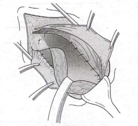
A trainee surgeon is performing his/her first cholecystectomy. The area marked with an asterisk in the diagram represents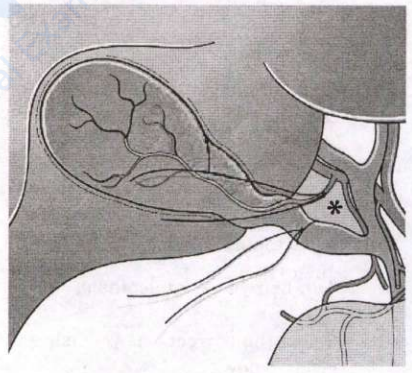
Calot's triangle
The asterisked area is Calot's triangle (cystohepatic triangle), bounded medially by the common hepatic duct, laterally by the cystic duct and superiorly by the inferior surface of the liver. It contains the cystic artery, the right hepatic artery, the cystic node of Lund and often accessory ducts. Clearing this triangle to obtain the critical view of safety, so that only two structures are seen entering the gallbladder, is the key step in preventing bile duct injury.
Rouviere's sulcus is a cleft on the right lobe marking the plane of the right portal pedicle, an extrahepatic landmark keeping dissection safely anterior, not a triangle. Bismuth lends his name to the classification of hilar cholangiocarcinoma and bile duct strictures, not to a triangle. The node of Lund (Mascagni node) is merely the sentinel node lying within Calot's triangle over the cystic artery, not the whole area.
Key point: Calot's triangle contains the cystic artery; divide no structure until the critical view of safety is achieved.
Similar Questions
Which one of the following statements regarding choledochal cyst is not correct?
Which one of the following is the most common benign liver lesion?
A 42-year-old lady underwent a laparoscopic cholecystectomy, which was uneventful other than some spillage of bile and gallstones during dissection. Postoperatively the patient was fine but she started complaining of right shoulder tip pain and developed high grade fever on the 3rd postoperative day. Her pulse was found to be 110/min and RR 20/min. The most likely diagnosis is
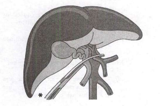
The manoeuvre (marked with an asterisk) shown in the following image is
Important Questions from Hepatobiliary & Pancreatic Surgery
- The Child-Turcotte-Pugh (CTP) score for quantifying the severity of chronic liver disease includes all variables EXCEPT:
'Chain of Lakes' appearance due to sacculation with intervening short strictures of pancreatic duct is seen on:
- Which one of the following statements is NOT correct regarding Pyogenic Liver Abscess?
- Which of the statements regarding Calot's triangle are correct?
1. Common hepatic duct forms the medial boundary of the Calot's triangle
2. Inferior surface of the right lobe of the liver forms the superior boundary of Calot's triangle
3. Right hepatic artery is usually found as a content of the Calot's triangle
4. Cystic duct and medial border of gall bladder forms the lateral border of Calot's triangle
Select the correct answer using the code given below: - CA 19-9 is a tumour marker used for the diagnosis of: