A 40-year-old female presents with symptoms of heat intolerance, excessive sweating and weight loss since 10-12 months. After clinical examination, the surgeon orders an investigation which is as shown in the image. The scan shown below is a/an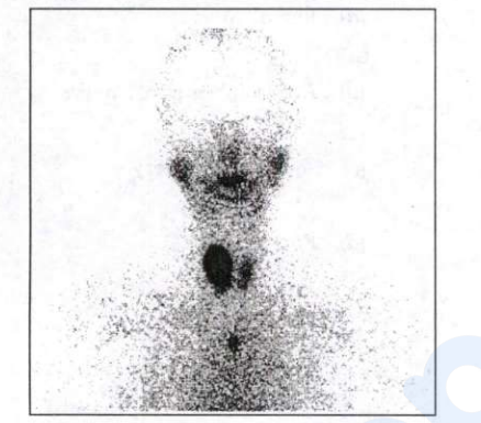
radionuclide scan
The correct answer is a radionuclide scan (thyroid scintigraphy). Heat intolerance, sweating and weight loss over 10-12 months indicate thyrotoxicosis, and the image is a functional isotope study using technetium-99m pertechnetate (half-life 6 hours, imaged at 20 minutes) or iodine-123/131. Its value is in finding the cause once TSH is suppressed: diffusely increased uptake in both lobes indicates Graves disease, a solitary hot nodule with a suppressed remaining gland indicates toxic adenoma, patchy multiple hot areas indicate toxic multinodular goitre, and near-absent uptake indicates thyroiditis or factitious thyrotoxicosis. It also detects retrosternal, lingual and ectopic thyroid tissue.
MRI and non-contrast CT are purely anatomical: they show size, retrosternal extension and tracheal compression but give no functional information, and iodinated contrast is best avoided in thyrotoxicosis. FDG-PET is an oncological tool for staging or detecting recurrent, iodine-negative thyroid carcinoma with a raised thyroglobulin, not for evaluating hyperthyroidism.
Key point: Function equals radionuclide scan; anatomy equals ultrasound, CT or MRI. A hot nodule is almost never malignant, whereas a cold nodule carries roughly 10-20 percent malignancy risk.
Similar Questions
An infant is brought to the clinic by the parents with the history of a swelling in the neck since birth. On examination, the swelling is found to be soft, partially compressible and brilliantly trans-illuminant. The most likely diagnosis is
A 29-year-old lady presents with a history of discharge from the nipple of the left breast since 1 month duration. Which one of the following is not warranted in the initial assessment of this patient?
A 45-year-old lady presents with a hard lump in the left breast. On examination, the lump is found to be 4 cm x 6 cm in size, not fixed to the skin, but fixed to the underlying pectoralis major muscle. The T stage of this is
Important Questions from Neck & Breast Surgery
- Medical management of thyrotoxic crisis includes all of the following EXCEPT:
- First line hormone therapy for post-menopausal woman with metastatic carcinoma breast is:
- All of the following are major subtypes of breast cancer based on Gene array analysis EXCEPT:
- Which of the following statements regarding lymphoedema following breast cancer treatment are correct?
1. Incidence has decreased due to rarely combined therapy of axillary LN dissection and
radiotherapy
2. Precipitating cause like LN metastasis is a major determinant
3. The condition is often painful
4. Oedematous limb is susceptible to bacterial infection
Select the correct answer using the code given below: - No increased relative risk of invasive breast carcinoma based on histopathological examination of benign breast tissue is for all of the following EXCEPT: