
The part of the human eye on which the image is formed is
retina
Understanding Image Formation in the Human Eye
The human eye is a remarkable organ that allows us to see the world around us. Light enters the eye and is focused onto a specific layer at the back, where it is converted into electrical signals that the brain interprets as images. Let's explore which part of the eye is responsible for forming this image.
Where the Image is Formed: The Retina
The crucial part of the human eye where the image is formed is the retina. The retina is a light-sensitive layer located at the back of the eye. It contains millions of photoreceptor cells, called rods and cones, which detect light. When light focused by the cornea and lens reaches the retina, these photoreceptor cells are stimulated. They convert the light energy into electrical signals.
These electrical signals are then processed by other neurons in the retina and transmitted to the brain via the optic nerve. The brain then interprets these signals to form the visual image we perceive. Therefore, the retina acts like the screen or film in a camera where the image is captured.
Analysis of Other Eye Parts and Their Roles
Let's look at the other options provided and understand their functions in the process of vision, clarifying why they are not the location where the image is formed:
- Pupil: The pupil is the opening in the center of the iris. Its size adjusts to control the amount of light entering the eye. It's like the aperture of a camera. Light passes *through* the pupil, but the image is not formed there.
- Cornea: The cornea is the transparent outer layer at the front of the eye. It is the first part that light encounters and is responsible for most of the focusing (refraction) of light entering the eye. It helps bend the light rays towards the lens, but the image is not formed on the cornea itself.
- Iris: The iris is the colored part of the eye surrounding the pupil. It acts like a diaphragm, controlling the size of the pupil and thus regulating the amount of light reaching the retina. It controls how much light enters, but it does not form the image.
Based on the functions of these parts, it is clear that the retina is where the light is focused and detected to begin the process of image formation.
Key Parts of the Human Eye and Vision
Here is a summary of the main parts involved in bringing light to the retina and their roles:
| Eye Part | Function | Role in Image Formation Location |
|---|---|---|
| Cornea | Transparent outer layer, primary light focusing | Helps focus light, but image is not formed here |
| Iris | Controls pupil size, regulates light amount | Regulates light entry, but image is not formed here |
| Pupil | Opening allowing light entry | Light passes through, but image is not formed here |
| Lens | Secondary light focusing, fine-tunes focus on retina | Helps focus light, but image is not formed here |
| Retina | Light-sensitive layer with photoreceptors | Image is formed here |
| Optic Nerve | Transmits electrical signals from retina to brain | Transmits signals, image is not formed here |
Conclusion on Image Formation in the Eye
The path of light involves passing through the cornea, pupil, and lens before being focused precisely onto the retina. It is on the retina that the light energy is converted into neural signals, marking the point where the initial step of image formation occurs before the signals are sent to the brain for interpretation.
Revision Table: Human Eye Anatomy and Function
| Anatomical Part | Primary Role |
|---|---|
| Cornea | Refraction of light |
| Iris | Controlling pupil size |
| Pupil | Allowing light entry |
| Lens | Adjustable focus (accommodation) |
| Retina | Light detection and signal generation |
| Optic Nerve | Signal transmission to brain |
| Vitreous Humor | Maintains eye shape |
| Aqueous Humor | Nourishes cornea & lens |
Additional Information on Eye and Vision
The process of vision is complex and involves multiple steps after the image is formed on the retina. The signals from the retina travel along the optic nerve to the visual cortex in the brain. The brain then processes these signals, combining information from both eyes and interpreting it to create our perception of the visual world, including depth, color, and movement. The fovea, a small area in the center of the retina, is responsible for sharp, detailed central vision because it has a high concentration of cone cells.
Conditions that affect the retina, such as macular degeneration or diabetic retinopathy, can significantly impair vision because they damage the layer where the image is initially captured and processed.
Similar Questions
Myopia is a defect in human vision where an image of a
Human eye can see objects at different distances with contrasting illuminations. This is due to
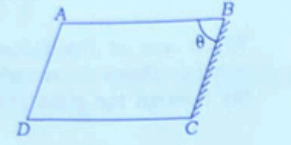
A glass slab of refractive index \(1\cdot5\) is cut in the shape of a parallelepiped \(ABCD\) as shown in the figure. Surface \(BC\) is polished to form a perfect reflector. Light is incident at surface \(AB\) from inside at an angle \(\pi/3\). Which one of the following is the correct angle \(ABC\) (\(\theta\)) such that the reflected light retraces its path?
- A periscope uses two plane mirrors inclined at \(45^\circ\). If the tube of the periscope is filled with a complete transparent liquid of refractive index \(1\cdot5\), then which one of the following is correct in respect of the image seen?
- Bottom of a tank containing water appears to be raised. It is due to
Statement I: Due to diffused or irregular reflection of light, a closed room gets light even if no direct sunlight falls inside the room.
Statement II: Irregular reflection, where the reflected rays are not parallel, does not follow the laws of reflection.Ramesh cannot see distinctly objects kept beyond 2 m. This defect can be corrected by using a lens of power
Light enters the eye through a thin membrane called
Colour vision in human eyes is the function of photoreceptor cells named
Cornea in human eye
Important Questions from Reflection and Refraction
Myopia is also known as ______.
Part of the eye which controls the light entering, is called as
Which defect of vision is corrected using cylindrical lenses in spectacles ?
Which of the following defects can be corrected by using a convex lens of appropriate power?
A person standing on the bank of a river observes a fish inside the water. He sees the fish_____.



