Point A in radiotherapy in carcinoma cervix marks the site for which of the following structures?
Crossing of ureter by uterine artery
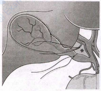
The correct answer is the crossing of the ureter by the uterine artery. In the Manchester system of intracavitary brachytherapy for carcinoma cervix, Point A lies 2 cm above the lateral vaginal fornix and 2 cm lateral to the cervical canal. It was defined to represent the paracervical tissue at the level where the uterine artery crosses over the ureter — the classic “water under the bridge” relation. This is the dose-limiting normal tissue, and the total Point A dose (about 80–85 Gy equivalent) governs both tumour control and the risk of ureteric stricture and fistula.
Obturator nodes describe Point B, which lies 5 cm lateral to the midline at the same height and represents the pelvic side-wall lymphatics. Inguinal nodes are irrelevant to cervical brachytherapy geometry — they drain the lower third of the vagina and vulva. The descending cervical artery is a branch of the uterine artery supplying cervix and upper vagina, but it is not the vessel defining the ureteric crossing used for Point A.
Key point: Point A equals 2 cm up and 2 cm lateral equals ureter crossed by the uterine artery; Point B equals 5 cm lateral equals obturator and pelvic side-wall nodes.
Similar Questions
The most common site for vulvar carcinoma is
Which one of the following states the diagnostic criteria for postmolar gestational trophoblastic neoplasia?
Important Questions from Neoplasms in Genital Tract
- Which one of the following is a protective factor for endometrial hyperplasia?
- Tumor marker of epithelial ovarian carcinoma is:
- The most common site of cervical cancer is:
- Which of the following are the vaccines for prevention of cervical cancer?
1. Cervarix
2. Gardasil
3. T-dap
4. Influenza
Select the correct answer using the code given below: - Consider the following statements regarding Carcinoma Cervix:
1. Clinical staging is done
2. Treatment if provided in stage I leads to survival rate of 80–90%
3. Surgery is preferred in young women with stage III disease
4. HPV virus is considered to be the causative agent
Which of the statements given above are correct?