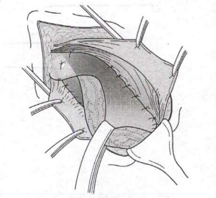
The typical ulcer as shown in the following diagram is seen in
tuberculosis
The correct answer is tuberculosis. The diagram shows an undermined (overhanging) edge, where the skin edge projects over a wider cavity so the base is broader than the surface opening. This happens when subcutaneous tissue is destroyed faster than the overlying skin, the classical behaviour of a tuberculous ulcer (and of an amoebic ulcer). Tuberculous ulcers are shallow with a pale wash-leather floor, thin bluish undermined edges and scanty seropurulent discharge, often over the neck from a collar-stud abscess of cervical nodes, caused by Mycobacterium tuberculosis. Biopsy shows caseating epithelioid granulomas; treatment is antitubercular therapy.
Syphilitic gummatous ulcers have a punched-out vertical edge. Squamous cell carcinoma gives an everted, rolled-out edge as proliferating tumour heaps over normal skin. Basal cell carcinoma (rodent ulcer) gives a rolled-in, beaded, pearly edge with telangiectasia.
Key point: Ulcer edges are a favourite one-liner: undermined = tuberculous, punched-out = syphilitic or trophic, everted = squamous carcinoma, beaded/rolled = basal cell carcinoma, sloping = healing.
Important Questions from Infections
- Which one of the following statements is NOT correct regarding Necrotising Soft Tissue infections?
- Which one of the following statements regarding Felon is NOT correct?
- All of the following are risk factors for an increased risk of wound infection EXCEPT:
- A 50 year old female presents to the emergency with pain, swelling and redness over the left foot following a trivial trauma 3 days back. On examina- tion, the swelling over the left foot is poorly localised; local tenderness and erythema are present and crepitus is absent; distal pulsations are palpable. The most likely clinical diagnosis is
- Mycetoma, a chronic, specific, granulomatous, destructive disease, involving the skin and subcutaneous tissue