
Question
Which one of the following microscopic techniques can be used to study the contour of proteins?
The correct answer is
AFM
Microscopic Technique for Protein Contour Analysis
To study the surface shape or contour of proteins at a microscopic level, a technique capable of high-resolution surface imaging is required.
Analyzing Microscopic Techniques
- Scanning Electron Microscopy (SEM): SEM provides high-resolution images of a sample's surface by scanning it with a focused beam of electrons. While useful for surface morphology, it often requires sample coating and may not offer the fine height-profile detail needed for precise protein contour, especially in liquid or near-native states.
- Transmission Electron Microscopy (TEM): TEM uses electrons that pass through a thin specimen to form an image. It excels at revealing internal structures but is less suited for imaging the external surface contour of proteins unless specific surface staining or shadowing techniques are used, which can alter the native structure.
- Atomic Force Microscopy (AFM): AFM is a type of Scanning Probe Microscopy. It uses a physical probe (tip) to scan across the surface of a sample. By detecting the forces between the tip and the sample, AFM creates a topographical map, directly measuring the surface height variations. This makes it ideal for determining the precise contour and shape of individual proteins with nanoscale resolution, often under physiological conditions.
- Confocal Microscopy: This is an optical technique used to obtain high-resolution 3D images of fluorescently labeled samples. It excels at imaging structures within thicker specimens (like cells) by rejecting out-of-focus light. However, it is limited by optical resolution and is not the primary method for detailed surface contour mapping of individual proteins.
Conclusion
Based on the capabilities of each technique, Atomic Force Microscopy (AFM) is the most suitable method for directly studying the detailed contour of proteins due to its high-resolution surface topography mapping.
The correct answer is Option C.
Was this answer helpful?
Important Questions from Protein Structure Folding Alpha Helix
- A 25 amino acid $\alpha$-helical protein is spanning the thickness of a mammalian cell membrane. Using the average dimensional parameters of a typical $\alpha$-helix, the thickness of the membrane will be ________ $\text{\AA}$ (rounded off to one decimal place).
The following diagram represents energy states of different protein folding steps, which includes-
1. Folded
2. Transition
3. Molten globule
4. Unfolded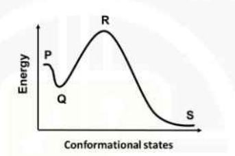
Find the correct match of folding steps (1 to 4) with the energy state (P to S) in the diagram.- In a $\alpha$-helix, the R-groups on the amino acid residues
- Which one of the following pairs of amino-acids in the protein has high propensity to take up the $\alpha$-helix conformation?
- Which one of the following closely defines 'Molten Globule' state of a protein?
Need Expert Advice?


