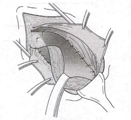
A 52-year-old carpenter presents with the history of a persistent non-healing ulcer on his lower alveolus. He is a smoker for last 30 years and also chews tobacco. What is the next most appropriate step in the management?
Edge biopsy of ulcer
The correct answer is edge biopsy of the ulcer. A persistent non-healing lower alveolar ulcer in a 52-year-old man who has smoked for 30 years and chews tobacco is oral squamous cell carcinoma until proved otherwise; tobacco nitrosamines, areca nut and alcohol act synergistically, and the gingivobuccal complex is the commonest oral subsite in India. Treatment depends entirely on histology, so tissue diagnosis must precede staging and therapy. Biopsy is taken from the edge, including a rim of normal mucosa, because the centre contains only necrotic slough and yields false negatives.
An X-ray of the mandible (or better, CT/MRI) assesses bone erosion and is part of staging after malignancy is confirmed; it cannot make the diagnosis. Local gum paint with astringent and analgesic is purely symptomatic and dangerously delays diagnosis. Systemic antibiotics would only suit an infective or dental-abscess ulcer and are inappropriate empirical treatment for a suspicious lesion.
Key point: Any oral ulcer failing to heal within 3 weeks in a tobacco user needs an edge biopsy; imaging is for staging and never substitutes for histopathology.
Similar Questions
A patient is undergoing a surgery for removal of a benign tumour from the parotid gland. Which one of the following structures should be preserved during this surgery?
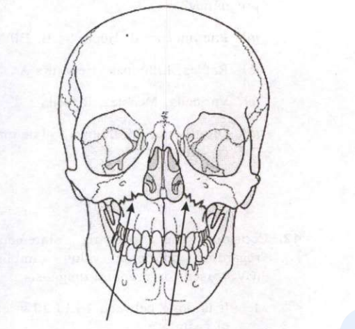
The type of fracture marked with arrow heads in the following figure is
Important Questions from Head
- A patient operated for a parotid gland tumour developed symptoms of sweating and erytherma (flushing) over the region of surgical excision while eating. The probable diagnosis is:
- Which of the statements regarding Salivary gland neoplasms are correct?
1. 80-90% of parotid tumors are malignant
2. 90% of sublingual gland tumors are malignant
3. 60-70% of submandibular gland tumors are benign
4. Parotid gland is most common site for salivary gland tumors
Select the correct answer using the code given below: - A young boy riding a motorcycle, met with a road traffic accident. On examination he had maxillofacial trauma with paraesthesia of the lower lip. Most likely underlying he has a:
- A 40-year-old smoker presents with soreness of tongue since last 2 months. On examination, it is found that the oral hygiene is poor and there is a white plaque over the lateral border of the tongue. What is the next step in the management of this patient?
- A 6-year-old boy has recurrent history of pain and swelling below his left ear, which generally lasts for 3-7 days and improves mildly after a course of antibiotics. Sialography shows punctate sialectasis. He should be treated by